IDOR develops Artificial Intelligence tools for the diagnosis of lung diseases

IDOR develops Artificial Intelligence tools for the diagnosis of lung diseases

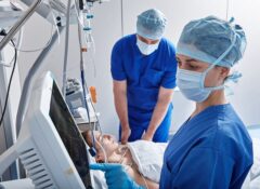
Scientists at the institute are creating customized tools to assist in interpreting images and reading radiological reports of lung exams. 

AI is undeniably one of the hottest topics in innovation. From banks and digital assistants to educational sectors, everyone is engaged in the quest for modernization and optimization that only this groundbreaking technology can deliver. In the healthcare sector, innovation and AI are particularly intertwined, promising significant advancements in medical care. 

Referring to this new technology as “intelligence” is more than just a poetic license. In reality, machine learning happens much like our own thought mechanisms: processing a vast amount of information to develop reasoning. The better the quality of information, the deeper our understanding of a particular subject. 

Setting aside our human subjectivity, we also diverge from AI in the amount of information that we’re able to process. While we may tire with a few hundred data points, machines adeptly process hundreds of thousands, maintaining a consistent level of accuracy without fatigue or distraction. 

In the healthcare AI race, radiology stands out as one of the most advanced fields. The precision of image analysis could significantly improve with tools that have thousands of references for various changes that may appear in exams. Rather than replacing radiologists, AI development in imaging optimizes the time for doctors and patients. These tools expedite image acquisition and diagnostic processes, offering an initial assessment to streamline healthcare professionals’ time. 

The potential for faster and more accurate diagnoses, coupled with the significant application of these tools in hospitals, is a key driver for the D’Or Institute for Research and Education (IDOR) in its AI research, specifically in thoracic radiology. 

AI Research at IDOR: 

AI development isn’t new at IDOR. The intensive care medicine research department at the institute focuses on analyzing large databases from intensive care units (ICUs) using machine learning. This approach enables computational systems to learn from extensive databases, optimizing their performance for specific tasks. 

In intensive care units, this optimization centers on identifying patterns that explain the evolution of different patient profiles, recognizing the impact of procedures, and guiding future care to enhance team performance and resource utilization. 

Identifying lung diseases through images: 

In the realm of image research, the developed tools aim to assist in diagnosing several lung diseases. This assistance unfolds in two investigative lines, utilizing different AI approaches. 

The first research line focuses on detecting, characterizing, and quantifying lung diseases such as pulmonary fibrosis and chronic obstructive pulmonary disease (COPD). An AI tool is gradually being trained to identify signs of different diseases in chest computed tomography images through a convolutional neural network (CNN), a technology mainly used for image processing and computer vision tasks. 

“At the moment, we are teaching the CNN various patterns and diseases. We have already used it in studies with COVID-19, and currently, we are training it with images of pulmonary emphysema. We are adding diseases, and our ultimate goal is to have a complete tool capable of characterizing a wide variety of diseases,” explains Dr. Rosana Rodrigues, a scientist at IDOR, a radiologist at Copa D’Or and Copa Star Rede D’Or hospitals and at the university hospital of the Federal University of Rio de Janeiro (UFRJ). 

According to Dr. Rosana Rodrigues, around 100,000 images of pulmonary emphysema, from over 300 tomographic exams, are being meticulously analyzed by her team. They mark the disease signs in each image, allowing the machine to identify patterns in its analyses, indicating the absence or presence of pulmonary emphysema, its subtypes, its extension, and stratifying patients by severity. The researcher details that these results only occur after many tests and checks by the team, a process that is repeated until the AI achieves a high degree of accuracy. 

Identifying lung diseases through reports: 

In addition to developing tools for reading lung radiological images, IDOR has also developed a solution for reading reports of chest computed tomography. One of the main goals of this research line is to identify potentially cancerous lung nodules in reports that were not performed for cancer screening, especially reports issued in emergency care. 

Rodrigues explains that the idea for the tool arose during the pandemic when many CT scans were performed to assess the need for hospitalization for COVID-19 patients. According to her, many of these images showed lung nodules that could be suspicious for cancer, but due to the urgency of the moment, these lesions were not always investigated. With social isolation measures, patients did not always return to receive their results. In this scenario, a machine capable of retrospectively assessing medical reports would be (and has been) very useful for identifying incidental findings of lung cancer in patients who were not being investigated for it. 

This work was detailed in a recent publication by Rodrigues’ team, composed of radiologists and developers who used another AI approach for this need. As the machine’s focus would be on characterizing nodules described in the exam reports, this tool would not need to interpret images but should be able to understand language variations present in the reports. With this in mind, Natural Language Processing (NLP) was the chosen AI resource for the development of the tool. 

The training process, however, was not very different from CNN, as more than 20,000 reports were used by scientists to promote the understanding of the machine. It underwent various supervised tests until achieving an incredible 98% accuracy in identifying malignant nodules. 

The sky is the limit 

Studies involving the use of AI in radiology are in full swing, and IDOR scientists view the expansion of these research efforts with enthusiasm. The CNN tool is in its final testing phases for broader clinical application, and researchers have already submitted their evaluation to the Brazilian Health Regulatory Agency (Anvisa). Meanwhile, the NLP tool has been used in various hospitals of Rede D’Or for the identification of patients who may have unnoticed lung cancer. “In AI, the sky is the limit; anything you think of is possible if you have a large database and a good developer,” comments Dr. Rosana Rodrigues. 

She further reveals that her group plans to develop similar tools for reading chest X-ray images and also plans to collaborate with other research areas at IDOR to investigate topics such as breast cancer and liver diseases. 

The improvement of AI tools for assistance in lung radiological examinations has much to contribute to healthcare. The technology has proven itself particularly relevant for the early identification of lung diseases, which have a higher likelihood of cure when treated in the early stages. 

Written by Maria Eduarda Ledo de Abreu.

More Content